Home
/ Muscle Chart Back / Amazon Com Muscles Male Poster 2 Views 24x36inch For Physical Fitness Working Out Muscular System Anatomical Chart Industrial Scientific
Muscle Chart Back / Amazon Com Muscles Male Poster 2 Views 24x36inch For Physical Fitness Working Out Muscular System Anatomical Chart Industrial Scientific
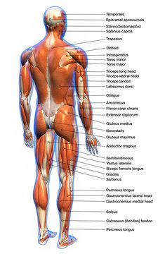
Muscle Chart Back / Amazon Com Muscles Male Poster 2 Views 24x36inch For Physical Fitness Working Out Muscular System Anatomical Chart Industrial Scientific. The trapezius and latissimus dorsi muscles connect the upper limb to the vertebral column. October 28, 2020 reading time: Claim your free copy of the client back care guide today. Build wide lats with this back building exercise. The rhomboid muscle is activated as you bring and squeeze your scapula or shoulder blades back and together.
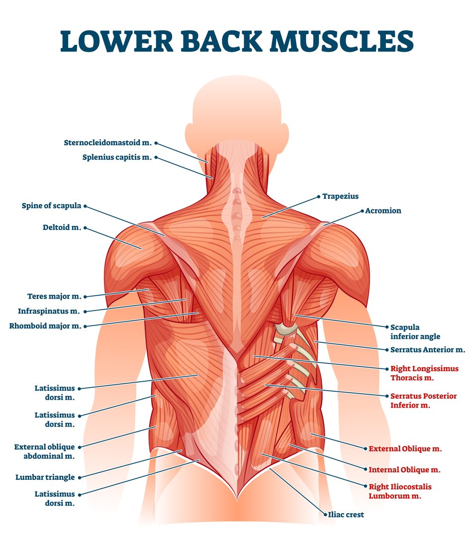
Muscle charts and stretching tips: We think this is the most useful anatomy picture that you need. Molly smith dipcnm, mbant • reviewer: The lordotic curve your lower back (lumbar spine) is the anatomic region between your lowest rib and the upper part of the buttock. This diagram shows which muscles in the lower back may be causing you pain.
Back Muscles Anatomy Chart 744 1140 Anatomy System Human Body Anatomy Diagram And Chart Images from anatomysystem.com The lordotic curve your lower back (lumbar spine) is the anatomic region between your lowest rib and the upper part of the buttock. The rhomboid muscle is activated as you bring and squeeze your scapula or shoulder blades back and together. While annoying, muscle knots — also called spasms or trigger points — can often be successfully treated with home remedies, trigger point release, stretching, strengthening and using good body mechanics. Muscles found in the superficial group include rhomboid major, rhomboid minor, levator scapulae, trapezius, latissimus dorsi. This procedure is one of the most powerful yet simple ways to treat muscle pain and discomfort. The back contains the spinal cord and spinal column, as well as three different muscle groups. Balance the weight of your head on top of your spine While muscles like the gluteals (in the thighs) are used any time we walk or climb a step, deep back muscles and abdominal muscles are usually not actively engaged during everyday activity.
Back muscle anatomy chart 12 photos of the back muscle anatomy chart back muscle anatomy chart, lower back muscle anatomy chart, human muscles, back muscle anatomy chart, lower back muscle anatomy chart.
Other muscles are small and cover much less space. Muscle charts and stretching tips: Muscle knots in your back can make everyday tasks, such as getting out of bed, painful and difficult. Back pain treatment many individuals will not need extensive treatment for back pain. Artery) p.134 accessory nerve p. Muscles connect to the vertebrae and bones via ligaments, flexible bands of fibrous tissue. The deltoid, teres major, teres minor, infraspinatus, supraspinatus (not shown) and subscapularis muscles (not shown) all extend from the scapula to the humerus and act on the shoulder joint. This curve, called lordosis, helps to: Some of these muscles are quite large and cover broad areas. The teres major is a small, yet important muscle within the back. While annoying, muscle knots — also called spasms or trigger points — can often be successfully treated with home remedies, trigger point release, stretching, strengthening and using good body mechanics. Muscles of back (trapezius, latissimus dorsi) Molly smith dipcnm, mbant • reviewer:
They lift and tilt head and lift or steady the shoulders. Back muscle anatomy chart 12 photos of the back muscle anatomy chart back muscle anatomy chart, lower back muscle anatomy chart, human muscles, back muscle anatomy chart, lower back muscle anatomy chart. 149 best skeletal muscle images in 2017 muscle tissue. Leaning back to straight vertical and all points in between. The back is the body region between the neck and the gluteal regions.
17 056 Best Back Muscles Anatomy Images Stock Photos Vectors Adobe Stock from t3.ftcdn.net We hope this picture anatomy of back muscles diagram can help you study and research. While annoying, muscle knots — also called spasms or trigger points — can often be successfully treated with home remedies, trigger point release, stretching, strengthening and using good body mechanics. Claim your free copy of the client back care guide today. Muscle anatomy neck 12 photos of the muscle anatomy neck dog neck muscle anatomy, front neck muscle anatomy, muscle anatomy neck, muscle anatomy of neck and shoulder, neck muscle anatomy chart, human muscles, dog neck muscle anatomy, front neck muscle anatomy, muscle anatomy neck, muscle anatomy of neck and. Artery) p.134 accessory nerve p. Back muscle diagram back muscles big back big back muscles big lats bodybuilding secrets major back muscles. Both the deltoid and the trapezius are firmly attached to the spine of the scapula. By the way, have you heard about the myth of.
We've created a free trigger point chart, which includes fybromyalgia treatment and reflexology information.
Muscle anatomy neck 12 photos of the muscle anatomy neck dog neck muscle anatomy, front neck muscle anatomy, muscle anatomy neck, muscle anatomy of neck and shoulder, neck muscle anatomy chart, human muscles, dog neck muscle anatomy, front neck muscle anatomy, muscle anatomy neck, muscle anatomy of neck and. Claim your free copy of the client back care guide today. Anatomynote.com found anatomy of back muscles diagram from plenty of anatomical pictures on the internet. Your clients will thank you for it! This procedure is one of the most powerful yet simple ways to treat muscle pain and discomfort. Muscles of the head and neck Most of the time, back muscle pain is diagnosed then treated with little more than a prescription of rest, painkillers and muscle relaxants. For more anatomy content please follow us and visit our website: Balance the weight of your head on top of your spine Muscle charts and stretching tips: Some of these muscles are quite large and cover broad areas. Facebook twitter google+ linkedin stumbleupon tumblr pinterest reddit vkontakte share via email print. The back's muscles start at the top of the back (named the cervical vertebrae) and go to the tailbone (also named the coccyx).
Intermediate back muscles and c. This procedure is one of the most powerful yet simple ways to treat muscle pain and discomfort. The two trapezius muscles extend from the backbone and base of the skull, across the back and shoulders to join the scapula and the clavicle. This diagram shows which muscles in the lower back may be causing you pain. 1) make midline incision along spines of vertebrae 2) extend from
Lower Back Muscle Anatomy And Low Back Pain from ix-cdn.b2e5.com Extrinsic and intrinsic.the back functions are many, such as to house and protect the spinal cord, hold the body and head upright, and adjust the movements of the upper and lower limbs. This website uses cookies to improve your experience while you navigate through the website. The lordotic curve your lower back (lumbar spine) is the anatomic region between your lowest rib and the upper part of the buttock. Out of these, the cookies that are categorized as necessary are stored on your browser as they are essential for the working of basic functionalities of the website. Muscle charts and stretching tips: Muscles of back (trapezius, latissimus dorsi) Anatomy chart courtesy of fcit the latissimus dorsi muscles (also known as the lats) are the largest muscles of the back. We think this is the most useful anatomy picture that you need.
Superficial (extrinsic) muscles of back.
Related posts of muscles of the lower back and hip diagram muscle anatomy neck. The trapezius and latissimus dorsi muscles connect the upper limb to the vertebral column. Superficial (extrinsic) muscles of back. This diagram shows which muscles in the lower back may be causing you pain. It comprises the vertebral column (spine) and two compartments of back muscles; The superficial group, the deep group, and the intermediate group. Anatomy chart courtesy of fcit the latissimus dorsi muscles (also known as the lats) are the largest muscles of the back. Balance the weight of your head on top of your spine Others, like sumo deadlifts, have been shown in emg studies—and in the trenches—to focus more on other muscle groups than the back. Muscles of the head and neck Back muscle anatomy chart 12 photos of the back muscle anatomy chart back muscle anatomy chart, lower back muscle anatomy chart, human muscles, back muscle anatomy chart, lower back muscle anatomy chart. The lordotic curve your lower back (lumbar spine) is the anatomic region between your lowest rib and the upper part of the buttock. This curve, called lordosis, helps to:
Share :
Post a Comment
for "Muscle Chart Back / Amazon Com Muscles Male Poster 2 Views 24x36inch For Physical Fitness Working Out Muscular System Anatomical Chart Industrial Scientific"


Post a Comment for "Muscle Chart Back / Amazon Com Muscles Male Poster 2 Views 24x36inch For Physical Fitness Working Out Muscular System Anatomical Chart Industrial Scientific"